Arteries and veins labeled model presents a comprehensive exploration of the circulatory system, providing an in-depth understanding of the structure, function, and clinical significance of arteries and veins. This guide delves into the anatomical features, physiological roles, and clinical implications of these vital vessels, offering a valuable resource for students, medical professionals, and anyone seeking to enhance their knowledge of human anatomy.
Through detailed diagrams, comparative tables, and engaging explanations, this guide illuminates the key differences between arteries and veins, highlighting their distinct roles in maintaining blood pressure, transporting oxygen and nutrients, and ensuring proper circulation throughout the body.
Arteries
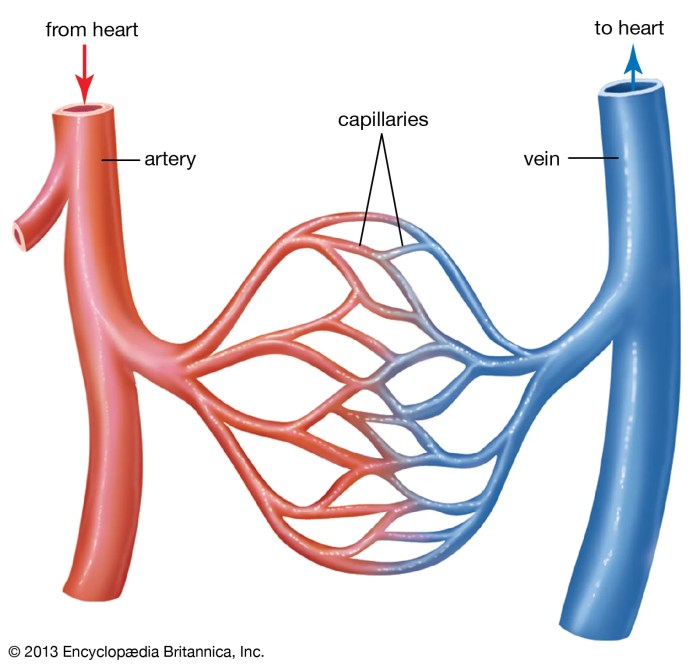
Arteries are blood vessels that carry oxygenated blood away from the heart to the rest of the body. They are responsible for maintaining blood pressure and circulation throughout the body.
The largest artery in the body is the aorta, which originates from the left ventricle of the heart. The aorta branches into smaller arteries that supply blood to the head, neck, arms, chest, abdomen, and legs.
Structure and Composition of Arterial Walls
Arterial walls are composed of three layers:
- Tunica intima:The innermost layer, which is lined with endothelial cells that prevent blood from clotting.
- Tunica media:The middle layer, which is composed of smooth muscle cells that control the diameter of the artery.
- Tunica adventitia:The outermost layer, which is composed of connective tissue that provides strength and support to the artery.
Role of Arteries in Maintaining Blood Pressure and Circulation
Arteries play a crucial role in maintaining blood pressure and circulation throughout the body. The elasticity of the arterial walls allows them to expand and contract, which helps to regulate blood flow and maintain a constant blood pressure.
When the heart contracts, it pumps blood into the arteries, which causes the arteries to expand. This expansion creates a pressure gradient that drives the blood flow throughout the body.
As the blood flows through the arteries, it loses pressure due to friction with the arterial walls. This loss of pressure causes the arteries to constrict, which helps to maintain a constant blood pressure.
Veins
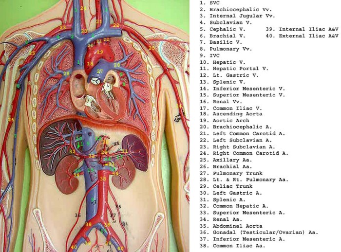
Veins are blood vessels that carry blood back to the heart. They are thin-walled and have valves to prevent backflow of blood. Veins are located throughout the body and are responsible for returning deoxygenated blood to the heart.
Structure and Composition of Vein Walls
Vein walls are composed of three layers:
- The tunica intima is the innermost layer and is lined with endothelial cells.
- The tunica media is the middle layer and is composed of smooth muscle cells.
- The tunica adventitia is the outermost layer and is composed of connective tissue.
The tunica intima is the most important layer of the vein wall as it prevents blood from leaking out of the vein. The tunica media helps to regulate blood flow by constricting or dilating the vein. The tunica adventitia provides support and protection for the vein.
Role of Veins in Returning Blood to the Heart
Veins play a crucial role in returning blood to the heart. They do this by creating a low-pressure system that allows blood to flow back to the heart. The valves in the veins prevent backflow of blood and help to maintain the low pressure.Veins
are an important part of the circulatory system and are essential for maintaining blood flow throughout the body.
Differences between Arteries and Veins
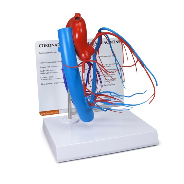
Arteries and veins are two types of blood vessels that form part of the circulatory system. They differ in their structure, function, blood flow, and oxygen content.
Structural Differences
- Arterieshave thicker, more muscular walls than veins. This is because they carry blood away from the heart at high pressure.
- Veinshave thinner, less muscular walls than arteries. This is because they carry blood back to the heart at low pressure.
- Arterieshave a narrower lumen (inner diameter) than veins. This is because they need to maintain a high pressure gradient to push blood through the body.
- Veinshave a wider lumen than arteries. This is because they need to accommodate the larger volume of blood returning to the heart.
Functional Differences
- Arteriescarry oxygenated blood away from the heart to the rest of the body.
- Veinscarry deoxygenated blood back to the heart.
Blood Flow Differences
- Arterieshave a high-pressure, pulsatile blood flow. This is because the heart pumps blood into the arteries with great force.
- Veinshave a low-pressure, continuous blood flow. This is because the heart does not pump blood into the veins; instead, the veins rely on the pressure gradient created by the arteries to push blood back to the heart.
Oxygen Content Differences
- Arteriescarry oxygenated blood, which is blood that has been enriched with oxygen in the lungs.
- Veinscarry deoxygenated blood, which is blood that has delivered its oxygen to the body’s tissues.
Implications for Circulatory Function
The differences between arteries and veins are essential for the proper functioning of the circulatory system. The thick, muscular walls of arteries allow them to withstand the high pressure of blood pumped from the heart. The thinner, less muscular walls of veins allow them to accommodate the larger volume of blood returning to the heart.
The high-pressure, pulsatile blood flow in arteries ensures that oxygenated blood is delivered to all parts of the body. The low-pressure, continuous blood flow in veins ensures that deoxygenated blood is returned to the heart.
Labeled Model
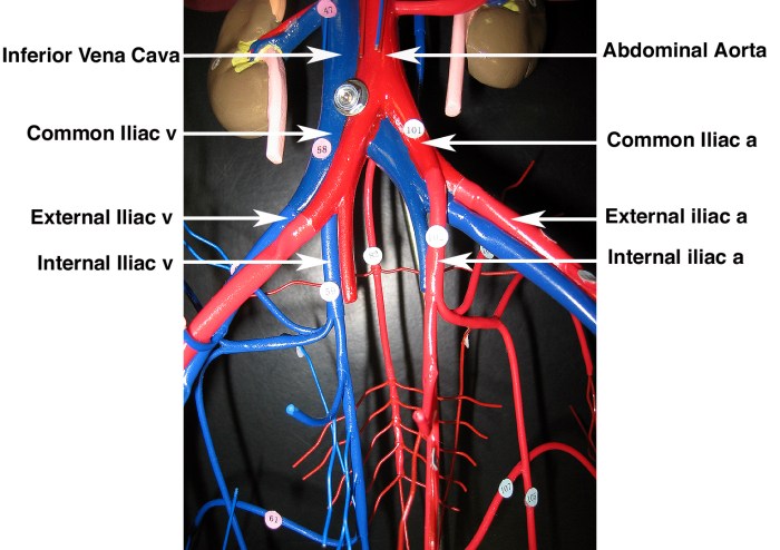
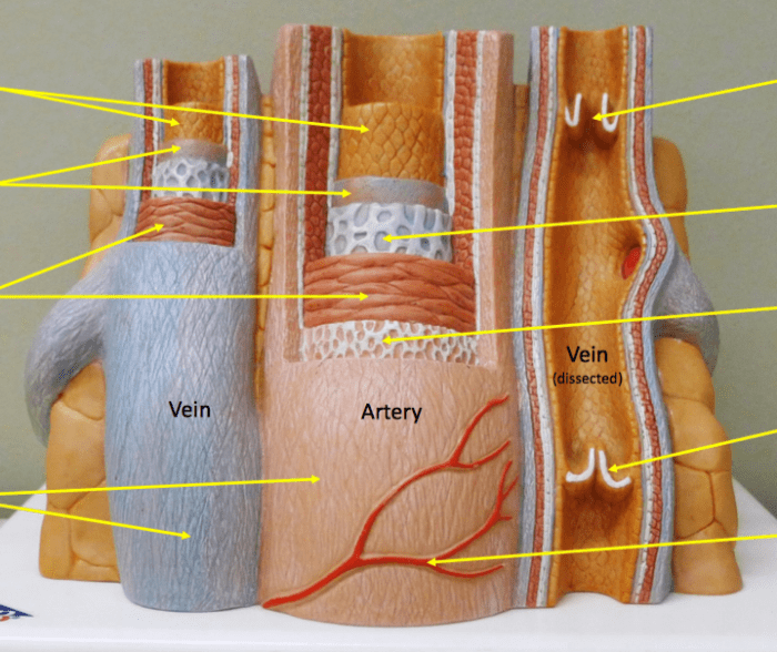
To effectively visualize and comprehend the structural differences between arteries and veins, a labeled model or diagram of their cross-sections can be designed. This model should meticulously annotate each anatomical feature, including the tunica intima, tunica media, and tunica adventitia.
The labeled model can serve as a valuable tool for understanding the distinct characteristics of arteries and veins. By examining the annotations, individuals can discern the variations in the thickness and composition of the three layers, as well as the presence or absence of valves.
Tunica Intima
The tunica intima is the innermost layer of both arteries and veins. In arteries, it is typically thicker and composed of a single layer of endothelial cells supported by a basement membrane and a layer of subendothelial connective tissue. In veins, the tunica intima is thinner and may contain multiple layers of endothelial cells.
Tunica Media, Arteries and veins labeled model
The tunica media is the middle layer of arteries and veins. In arteries, it is composed of smooth muscle cells arranged in concentric layers. In veins, the tunica media is thinner and contains fewer smooth muscle cells, which are often arranged in a more irregular pattern.
Tunica Adventitia
The tunica adventitia is the outermost layer of arteries and veins. In arteries, it is composed of connective tissue and contains collagen fibers, elastic fibers, and fibroblasts. In veins, the tunica adventitia is thinner and contains more connective tissue and fewer elastic fibers.
Clinical Significance
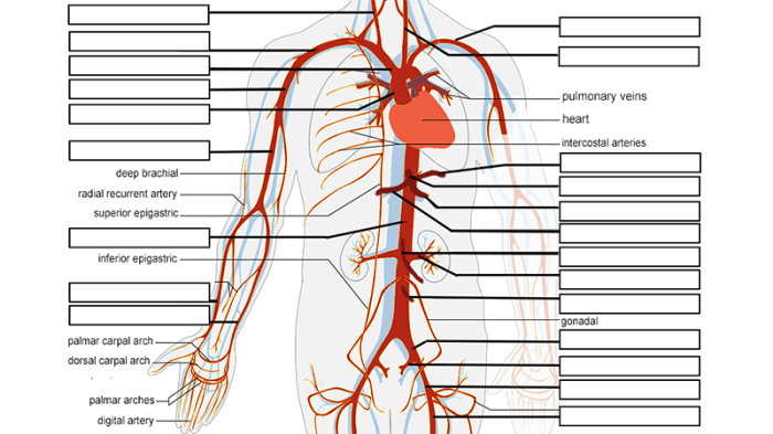
Understanding the anatomy and function of arteries and veins is crucial for medical professionals due to their central role in the circulatory system. A thorough grasp of these vessels aids in the diagnosis and treatment of various medical conditions and surgical interventions.
Medical Conditions and Procedures
- Arterial Hypertension (High Blood Pressure):Occurs when blood pressure in arteries is abnormally elevated, putting strain on the vessels and increasing the risk of cardiovascular events like heart attack or stroke.
- Arteriosclerosis:A condition where arteries become thickened and hardened due to plaque buildup, restricting blood flow and increasing the risk of blockages or clots.
- Varicose Veins:Enlarged, twisted veins that often occur in the legs, causing discomfort and potential complications like blood clots or skin ulcers.
- Deep Vein Thrombosis (DVT):A serious condition where blood clots form in deep veins, typically in the legs, posing a risk of pulmonary embolism if the clot travels to the lungs.
Diagnosis and Surgical Interventions
Understanding arterial and venous anatomy is essential for accurate diagnosis and effective surgical interventions:
- Angiography:A medical imaging technique that uses X-rays and contrast dye to visualize arteries, allowing for the detection of blockages, narrowing, or abnormalities.
- Venography:Similar to angiography, but used to visualize veins, aiding in the diagnosis of conditions like DVT or varicose veins.
- Arterial Bypass Surgery:A surgical procedure to create a new pathway for blood flow around a blocked or narrowed artery, restoring circulation and preventing tissue damage.
- Endovenous Laser Treatment:A minimally invasive procedure that uses laser energy to seal off varicose veins, improving blood flow and reducing symptoms.
Q&A: Arteries And Veins Labeled Model
What is the main difference between arteries and veins?
Arteries carry oxygenated blood away from the heart to the body’s tissues, while veins carry deoxygenated blood back to the heart.
Why are arteries thicker than veins?
Arteries have thicker walls to withstand the higher pressure of oxygenated blood being pumped from the heart.
What is the role of valves in veins?
Valves in veins prevent blood from flowing backward, ensuring that blood flows towards the heart.
